Scientists develop 5D technique to identify tell-tale signs of disease from cell phone pictures
16 Jan 2017
A new 5D technique developed by scientists will allow doctors to analyse images to quickly identify tell-tale signs of diseases from pictures taken using cell phones, PTI reported.
"Hyper-Spectral Phasor" analysis, or HySP, as the techniques is called is much faster and far less expensive than current techniques, and might be useful for diagnosing and monitoring diseases by using cell phone images, according to researchers.
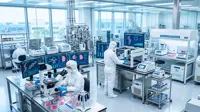
With the new imaging technology, researchers at the University of Southern California (USC) in the US had used fluorescent imaging to locate proteins and other molecules in cells and tissues.
In the technique, molecules are tagged with dyes that glow under certain kinds of light - the same principle behind so-called "black light" images.
Fluorescent imaging could help scientists understand which molecules were produced in large amounts in cancer or other diseases, information that might be useful in diagnosis or in identifying possible targets for therapeutic drugs.
Looking at just one or two molecules in cell or tissue samples was fairly straightforward, however, it did not provide a clear picture of how those molecules were behaving in the real world.
"Biological research is moving toward complex systems that extend across multiple dimensions, the interaction of multiple elements over time," said Francesco Cutrale, a postdoctoral fellow at the USC.
"By looking at multiple targets, or watching targets move over time, we can get a much better view of what's actually happening within complex living systems," Cutrale said.
The researchers had tested the concept on zebra fish so far and in the future the team planned to test HySP on humans, specially members of the military who had been exposed to chemicals and materials damaging to their lungs while in combat. A tiny probe with a flourescent light would be lowered into their lungs to capture images of the surrounding tissues. These pictures would be compared with those of healthy lungs, and researchers hope they would be able to spot a difference.
If successful, the researchers hope the technology would be useful in practice to diagnose lung disease. They note that their technology could be used in cell phone cameras as well, users could take a picture of a lesion and a doctor could tell you if it was potentially cancerous.