Study finds how pancreatic cancer spreads to liver
28 May 2015
An international team led by Weill Cornell Medical College investigators has illuminated the precise molecular steps that enable pancreatic cancer to spread to the liver – the event that makes the most common form of the disease lethal.
 By understanding this process, investigators say their discovery can lead to targeted treatments that delay metastasis and could offer clinicians a new biomarker to test for the earliest signs of pancreatic cancer.
The study, published 18 May in Nature Cell Biology, focuses on the role of small, spherical tumour-secreted packages, called exosomes, which contain tumour-derived proteins, in preparing a liver micro-environment fertile for pancreatic cancer metastasis.
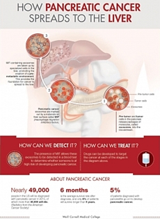
Nearly 49,000 people in the United States will be diagnosed with pancreatic cancer, and more than 40,000 of them will succumb to the disease, according to estimates from the American Cancer Society. Pancreatic cancers are among the most lethal – only 6 per cent of patients survive five years after diagnosis; the median survival rate is just six months.
''What makes this cancer so lethal is that patients don't generally become symptomatic – and as such aren't diagnosed – until the cancer is very advanced and treatment options are limited,'' said senior author Dr. David Lyden, the Stavros S. Niarchos Professor in Pediatric Cardiology and a professor of pediatrics at Weill Cornell.
In the study, the investigators re-created the environment for pancreatic cancer using mouse models and discovered that exosomes were finding their way to the liver during the cancer's earliest stages.
Once in the liver, the exosomes were taken up by resident immune cells, called Kupffer cells. This process changed the Kupffer cells' gene expression and protein composition, and educated them to produce a powerful protein. This protein, in turn, affected the behavior of a group of cells, inducing liver fibrosis – an overly exuberant wound healing process that can interfere with normal liver function and creates a microenvironment auspicious for tumor seeding and growth.
When investigating how exosomes exerted these effects on liver cells, Lyden and his team found that pancreatic cancer exosomes contain a protein called macrophage migration inhibitory factor (MIF). When the investigators eliminated MIF from exosomes, they noticed that they had prevented the creation of a fibrotic, tumor-supporting environment in the liver.
''In mouse models of pancreatic cancer progression, exosomes containing MIF are released in circulation prior to the onset of a recognized pancreatic carcinoma and can 'educate' the liver, inducing fibrosis,'' says first author Dr. Bruno Costa Silva, an instructor of cell and developmental biology in pediatrics at Weill Cornell. ''Our findings suggest that a microenvironment ripe for metastasis is generated at an earlier stage of the disease than previously recognized.''
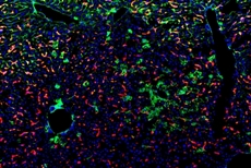 | |
| MIF-expressing pancreatic cancer exosomes induce/prepare liver metastasis. Pancreatic cancer exosomes induce liver fibrosis (fibronectin, green) and immune cells (macrophages, red) accumulate / Image: Dr. Bruno Costa da Silva and Dr. David Lyden |
''Disrupting just one part of the process at any point of the circuit decreased metastasis, a discovery that could lead to the development of multi-targeted therapies that could prolong patients' lives,'' says Lyden, who also has appointments in the Sandra and Edward Meyer Cancer Center and the Gale and Ira Drukier Institute for Children's Health. Lyden and his team conduct their research in the Children's Cancer and Blood Foundation labs at Weill Cornell.
Lyden and his team also found that MIF is highly expressed in exosomes circulating in patients who have advanced pancreatic cancer. When they examined pancreatic cancer blood samples, the scientists discovered that exosomal MIF was much higher in patients who went on to develop liver metastasis than in those who escaped it.
They say this protein signature could be used to predict which patients would go on to develop liver metastatic disease. These discoveries were made possible by an international collaboration among researchers at Weill Cornell, Memorial Sloan Kettering Cancer Center, University of Nebraska Medical Center, University of Pennsylvania and Oslo University Hospital.
Since five per cent of patients diagnosed with pancreatitis – a disease characterised by inflammation – go on to develop pancreatic cancer, the investigators believe MIF also could serve as a biomarker for clinicians to monitor disease progression.
Lyden and his team are testing whether measuring MIF levels in exosomes isolated from patients' blood can accurately estimate the risk of pancreatic cancer in patients with nonmalignant pancreatic lesions. This type of ''liquid biopsy'' could allow the clinicians to initiate treatments, such as surgical resection, earlier in patients at risk, preventing disease progression.