New type of fluorescent camera for blood diagnostics, brain mapping developed
By By Matthew Chin | 28 Sep 2013
Fluorescence imaging is the most widely used method for analysing the molecular composition of biological specimens. Target molecules, when they are present, can be "tagged" with a fluorescent label and made visible.
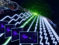 |
| Artist's conception of an action potential propagating along a neuron, as imaged by the FIRE camera |
This highly sensitive technique, which is used in screening blood for cancer cells and studying biochemical reactions, is very good at detecting molecules present in extremely low concentrations.
However, because of the very small amount of light produced by fluorescent molecules, current state-of-the-art camera technologies must image samples slowly in order to collect enough photons to generate an image.
Further, these cameras typically read out individual pixels one at a time to create an image. These two factors limit the speed at which fluorescence imaging can be performed.
Inspired by how wireless communication networks use multiple radio frequencies to communicate with multiple users, researchers from the University of California Los Angeles (UCLA) Henry Samueli School of Engineering and Applied Science have developed a new high-speed microscopy technique that is an order of magnitude faster than current fluorescence-imaging technologies.
The research, published online in the peer-reviewed journal Nature Photonics, was led by principal investigator Bahram Jalali, a professor of electrical engineering and bioengineering who holds UCLA's Northrop Grumman Opto-Electronic Chair in Electrical Engineering; lead author Eric Diebold, a UCLA postdoctoral scholar in electrical engineering; and Brandon Buckley, a UCLA Ph.D. student in physics. The team also included Daniel R. Gossett, a recent UCLA bioengineering Ph.D. graduate.
"Telecommunication systems routinely generate and analyse data at billions of bits per second," Jalali said. "Interestingly, the amount of data that needs to be captured and processed is the same in many biomedical imaging applications. This demonstration is a perfect example of how technology from the field of communications can be used in a new way to solve problems in other areas of science."
While current cameras form images of fluorescent samples pixel by pixel, the camera created by the UCLA team forms an image by reading an entire row of pixels at once. It does this by encoding the fluorescence from each pixel on a different radio frequency; in this way, it can form images at a rate approximately 10 times faster than current state-of-the-art technologies.
"We've figured out a way to encode the image pixels in the frequency domain such that we can use a single-pixel detector as a multi-pixel detector," Diebold said. "The signal from a single-pixel detector can be read and saved much more quickly than a multi-pixel detector, such as those in conventional cameras.
It uses principles similar to the way that many TV channels are carried on a single wire that goes into your house, or how multiple computers can communicate with a single wireless Internet router at the same time. Using that as inspiration, we treated each pixel on the image as a different channel."
With the new technique, a laser beam is first split into two beams, with one of the beams changed slightly in frequency. The two beams then combine again on the sample that has been labeled with fluorescent molecules.
As they interfere at the sample, the frequency difference between the two beams encodes the fluorescence from each pixel, allowing the camera can detect an entire row of pixels at one time. The researchers call the new technique "fluorescence imaging using radiofrequency-tagged emission," or FIRE.
The team says the technique can be applied in flow cytometry, which analyzes cells one by one in a flowing fluid. Unlike current imaging flow cytometers, the FIRE technique can keep up with conventional flow-cytometry speeds and image 50,000 cells per second. While most flow cytometers take only a single-point estimate of a cell's fluorescence, FIRE can create an entire high-resolution, multicolor image of each cell, providing significantly more information to a biologist or clinician.
"This information could be the difference between detecting a rare cancer cell in a blood sample or missing it completely," Diebold said. "We're able to capture images of cells that are blur-free as they flow by at a speed around 100 times faster than state-of-the-art imaging flow cytometers," Diebold said. "This technique really gives you a speed advantage that puts you into a new performance regime relative to current technologies."
The researchers say the technique could also be applied in vivo - for example, in neuroscience research to view neural activity in the brain, making a much larger field of view available to analyze.
"In biology, there are a lot of phenomena that happen at the sub-millisecond time scales, like when the brain fires action potentials with pulses that last around one millisecond" Diebold said. "To resolve a millisecond event, you want to sample that at least twice in that one millisecond, so you need half-millisecond time resolution from your imager.
"A lot these higher-speed fluorescence imaging cameras can get close to single-millisecond time resolution but only over a really small field of view," he said. "They read out pixels serially, one after the other. Our FIRE method gets us well beyond that, as we can simultaneously analyze more than 200 pixels in a row, making a much bigger field of view available at one time."
Jalali also holds a faculty appointment in department of surgery at the David Geffen School of Medicine at UCLA and is a member of the California NanoSystems Institute at UCLA.
The research was partially funded by the Broad Stem Cell Research Center at UCLA.
The UCLA Henry Samueli School of Engineering and Applied Science, established in 1945