Researchers image glycans on embryonic cells hours after fertilisation
10 Jul 2010
Researchers with the US Department of Energy's Lawrence Berkeley National Laboratory (Berkeley Lab) and the University of California (UC), Berkeley, have successfully attached imaging probes to glycans - the sugar molecules that are abundant on the surfaces of living cells - in the embryos of zebrafish less than seven hours after fertilization. Glycans are key regulators of the processes that guide cell development, and zebrafish are a top vertebrate model organism of embryogenesis. This new technique enables scientists to study the physiological changes cells undergo during embryogenesis without invading and doing damage to the embryos.
The team of researchers led by Carolyn Bertozzi, a Berkeley Lab-UC Berkeley chemical biologist and leading authority on glycobiology, used a combination of glycan metabolic labeling and copper-free click chemistry to record the earliest images ever of glycan activity on embryonic cells. The images were obtained during a stage of development in which many of the cells were still in the ''multipotent stem cell'' state, meaning they had yet to differentiate into specific tissue types.
''We know from earlier studies of developmental biology that glycan structures can change a lot during the early stages of embryogenesis,'' Bertozzi says. ''With this new technology, we hope to witness some of those changes in the glycome in real time and to understand better how cell surface glycans might contribute to the decisions that stem cells make about their destiny.''
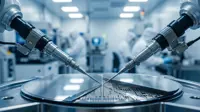
A research team led by Carolyn Bertozzi (seated) and including (from left) Scott Laughlin, Sharon Amacher and Karen Dehnert successfully attached imaging probes to glycans in the embryos of zebrafish less than seven hours after fertilization, the earliest recordings ever of of glycan activity on embryonic cells. (Photo by Roy Kaltschmidt, Berkeley Lab Public Affairs).
A research team led by Carolyn Bertozzi (seated) and including (from left) Scott Laughlin, Sharon Amacher and Karen Dehnert successfully attached imaging probes to glycans in the embryos of zebrafish less than seven hours after fertilization, the earliest recordings ever of of glycan activity on embryonic cells. (Photo by Roy Kaltschmidt, Berkeley Lab Public Affairs).
Bertozzi is the director of Berkeley Lab's Molecular Foundry, a faculty scientist with Berkeley Lab's Materials Sciences and Physical Biosciences Divisions, and the T.Z. and Irmgard Chu Distinguished Professor of Chemistry as well as a professor of Molecular and Cell Biology at UC Berkeley, and an investigator with the Howard Hughes Medical Institute (HHMI).