IBM and ETH scientists advance supercomputing simulations to improve diagnosis of osteoporosis
02 July 2008
Using an IBM Blue Gene/L supercomputer, scientists of the ETH Zurich (Swiss Federal Institute of Technology) and the IBM Zurich Research Laboratory demonstrated the most extensive simulation yet of real human bone structures, providing doctors with a ''high-definition'' view of fragility or strength of bones they never had before. This achievement could lead to better clinical tools to improve the diagnosis and treatment of osteoporosis, a widespread disease that affects 1 in 3 women and 1 in 5 men over the age of 50.1
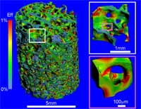 |
| Photo: ETH Zurich, IBM Research Researchers of ETH Zurich and the IBM Zurich Research Laboratory have demonstrated the most extensive simulations of a real human bone structure ever achieved. In order to analyze bone strength, the researchers used massively parallel simulations to obtain a ''heat map'' of strain, which changes with the load applied to the bone. The image shows the effective strain on a 5 by 5 by 5 mm human vertebra specimen under a load corresponding to the person's weight when standing. The areas in blue show stronger parts of the bone whereas weaker parts are shown in red. The simulations are based on a high-resolution scan (6 microns) using peripheral quantitative computer tomography (pQCT). The researchers created a very detailed mathematical model that required more that 400 million elements-called "voxels"-in order to capture faithfully the intricate bone microstructure. In the next step, researchers computed the strain on the bone by applying different loads that simulate real-life conditions. The specimen in this image shows a healthy human bone. |
Osteoporosis is the most widespread bone disease worldwide, affecting 75 million people in the US, Europe and Japan alone, and causing health costs second only to those associated with cardiovascular diseases. Literally ''porous bone'', this disease is characterized by loss of bone density, resulting in a high risk of fractures, and is a major cause of pain, disability and death in older persons.Unfortunately, in many cases, osteoporosis is not diagnosed until a fracture has occurred, but by then the disease is already in an advanced stage, requiring implants or surgical plates to treat or prevent further fractures (source: Prevention and Management of Osteoporosis, WHO Technical Report Series, No. 921.).
Today, osteoporosis is diagnosed by measuring bone mass and density using specialized x-ray or computer tomography techniques?a highly empirical process.
Studies have shown, however, that bone mass measurements are only a moderately accurate way to determine the strength of the bone because bones are not solid structures. Inside the compact outer shell, bones have a sponge-like center. This complex microstructure accounts for the bone's capability to bear loads and therefore represents a better indicator of a bone's true strength.
Aiming for an accurate, powerful and fast method to automate the analysis of bone strength, scientists of the Departments of Mechanical and Process Engineering and Computer Science at ETH Zurich teamed up with supercomputing experts of IBM's Zurich Research Laboratory. The breakthrough method they developed combines density measurements with a large-scale mechanical analysis of the inner-bone microstructure.
