Prototype NIST / CU bioreactor evaluates engineered tissue while creating it
05 May 2012
Researchers at the National Institute of Standards and Technology (NIST) have developed a prototype bioreactor - a device for culturing cells to create engineered tissues - that both stimulates and evaluates tissue as it grows, mimicking natural processes while eliminating the need to stop periodically to cut up samples for analysis. Tissue created this way might someday be used to replace, for example, damaged or diseased cartilage in the knee and hip.
 Conventional methods for evaluating the development and properties of engineered tissue are time-consuming, destructive and need to be repeated many times. By using ultrasound to monitor tissue during processing without destroying it, the novel bioreactor could be a faster and less expensive alternative.
"Most bioreactors don't do any type of nondestructive evaluation," says NIST post-doctoral researcher Jenni Popp, first author of a new paper about the instrument. "Having some sort of ongoing evaluation of the developing tissue is definitely novel."
Cartilage is smooth connective tissue that supports joint motion. Natural cartilage is created by specialised cells that generate large amounts of structural proteins to weave a tough support material called extracellular matrix. Lacking blood vessels, cartilage has limited capability to heal from arthritis, sports injuries or other defects.
Damage can be treated with drugs or joint replacement but results can be imperfect. Engineered tissue is used in some medical treatments but is not yet a routine alternative to metal or plastic joint replacements. The NIST bioreactor gives researchers a noninvasive way to monitor important structural changes in developing tissue.
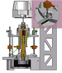
The NIST / CU bioreactor can fit inside a standard incubator, which controls temperature and acidity in the growth environment. The bioreactor applies force to stimulate five small cubes of cartilage cells embedded in water-based gels. The mechanical force mimics the natural stimuli needed for the cells to create matrix proteins and develop the structure and properties of real cartilage.
Ultrasound techniques monitor tissue changes over time, while a digital video microscope takes images.
Preliminary studies indicate the bioreactor both stimulates and monitors development of cells, matrix content and scaffolds to make three-dimensional engineered cartilage. The cell-laden gels were stimulated twice daily for an hour. Sulfated glycosaminoglycan (sGAG) - which combines with fibrous proteins to form the extracellular matrix-increased significantly after seven days. This structural change was detected by a significant decrease in ultrasound signals after seven days.
The research described in the new paper was performed at and led by NIST. The bioreactor is a collaborative project with several co-authors from the University of Colorado Boulder (CU) Department of Chemical and Biological Engineering.
NIST and CU researchers continue to develop ultrasonic measurement methods and plan to conduct longer experiments. The bioreactor is also being used by other academic researchers as a tool for validating mathematical models of biokinetics, the study of growth and movement in developing tissue.